Protocol
Construction of GFP plasmid
Transformation
Materials and reagents
- 1. Competent cell (Escherichia coli DH5α)
- 2. LB plate (0.5% yeast extract, 1% tryptone, 1% NaCl, 1.5% agar) CP+)
- 3. Plasmid: pSB1C3 ‐ BBa_I746909 (superfolder GFP driven by T7-promoter)
- 4. Super optimal broth (SOB)
Procedure
- 1. Take out the competent cells from ‐80°C refrigerator
- 2. Thaw the cells on ice for 5 minutes, and prewarm SOB at 37°C
- 3. Add 1 μL plasmid DNA into the competent cells, and mix gently by pipetting
- 4. Put on ice for 10 minutes
- 5. Heat shock at 37°C for 3 minutes
- 6. Put on ice for 2 minutes
- 7. Add 150 μL 37°C SOB into the mixture
- 8. Place the tube in a 37°C incubator for 4 hours
- 9. Spread the cells onto the LB plate(Cp+)
- 10. Incubate the plate at 37°C with shaking for 14‐16 hours
Mini‐preparation of plasmid
Materials and reagents
Procedures
- 1. Add 2 ml LB broth into 15 ml tube
- 2. Add 2 μl Cp into the tube
- 3. Use the tip to scrape up a colony on LB plate(Cp+), and detach the tip into the tube
- 4. Put the tube into a 37°C shaking incubator for 8 hours
Midi‐preparation of plasmid
Materials and reagents
Procedures
- 1. Add 100 ml LB broth into a flask
- 2. Add 100 μl Cp into a flask
- 3. Transfer 500 μl broth of mini‐preparation into a flask
- 4. Put the flask into a 37°C shaking incubator for 15 hours
Extraction of plasmid
Materials and reagents
- plasmid extraction kit (invitrogen)
Procedure
- 1. Divide 100 ml broth in the flask into two 50 ml tubes
- 2. Centrifuge two tubes for 10 minutes at 4000 xg , 4°C and remove the supernatant
- 3. Add 10 ml EQ buffer to wash column
- 4. Add 4 ml mp1 to one tube and vortex to suspend bacteria clump, and transfer suspension to another tube and vortex
- 5. Add 4 ml mp2 , invert the tube and stand for 5 minutes
- 6. Add 4 ml mp3 and invert the tube until precipitate appears
- 7. Centrifuge the tube for 10 minutes at 4000 xg , 4°C
- 8. Place filter paper on column and add 1 ml MilliQ water to wet the paper.
- 9. Transfer supernatant and let it go through the filter paper into column
- 10. Add 10 ml wash buffer to wash column twice
- 11. Move the column on 15 ml tube, add 5 ml elution buffer to column and collect the flow‐through
- 12. Add 0.7 volume of isopropanol to the tube and invert several times
- 13. Centrifuge the tube for 30 minutes at 9000 xg , 4°C and remove the supernatant
- 14. Add 1 ml ethanol and suspend the pellet, and transfer to a ppendorf
- 15. Centrifuge the tube for 5 minutes at 12000 xg , 4°C and remove the supernatant
- 16. Open the eppendorf and air‐dry for 20 minutes
- 17. Add 50μl TE buffer
- 18. Use Nanodrop to measure plasmid DNA concentration and record the concentration, 260/280, 260/230
Restriction digestion of plasmid
Materials and reagents
- 1. dH2O
- 2. Restriction enzymes EcoRI and PstI
- 3. 10X buffer
- 4. Plasmid: pSB1C3 ‐ BBa_I746909 (superfolder GFP driven by T7-promoter)
Procedures
- 1. Use restriction enzyme EcoRI to digest the plasmid
- Prepare the mixture for restriction digestion following the table list
| reagents |
volume(μl) |
| Restriction enzyme EcoRI |
0.4 |
| dH2O |
16.6 |
| 10X buffer( EcoRI buffer) |
2.0 |
| Plasmid |
1.0 |
| total volume |
20 |
2. Use two restriction enzyme EcoRI and PstI to digest the plasmid- Prepare the mixture for restriction digestion following the table list
| reagents |
volume(μl) |
| Restriction enzyme EcoRI |
0.4 |
| Restriction enzyme PstI |
0.4 |
| MilliQ water |
16.2 |
| 10X buffer( EcoRI buffer) |
2.0 |
| Plasmid |
1.0 |
total volume |
20 |
3. Put the mixture at 37°C for 1 hour
Garose gel electrophoresis
Materials and reagents
- 1. Agarose
- 2. 1X TAE buffer
- 3. 10X loading dye
- 4. 1 kb marker
- 5. Ethidium bromide (EtBr)
Procedure
- 1. Gel preparation
- (1) Prepare 8‐well 1.0% agarose gel with 1X TAE buffer in a total volume of 25 ml
- (2) Use microwave to heat up the solution until boiling and place the solution in
water bath to cool down
- 2. Sample loading
- (1) Digested plasmid samples are mixed with the 10X loading dye.
- (2) Load all samples and markers into well
- 3. Gel running and staining
- Run the gel at s constant voltage of 100V for 20 min, and then stain the gel with
Ethidium bromide for 10min for UV visualization.
DNA folding reaction
Preparation of staple mixtures and scaffolds
NanoNeedle consists of 2 scaffolds, p7249 (M13mp18) and p8064, 427 staples, and 9 staples with aptamers. Three component were respectively ordered from tilibit, MDBio, and IDT.
- 1. 436 staple tubes are dissolved with MilliQ water according to information provided by the company, and each concentration is 100 µM.
- 2. Then 2.5ul of each staples are added and mixed together, and final concentration of each staple is 100nM.
- 3. We divide each scaffold tubes into 5 tubes for continued experiment, and each concentration is 100nM.
preparation of folding buffer and MgCl2 solution
- 1. We prepare 10X folding buffer containing 50 mM TRIS 8.0, and 10 mM EDTA from 1M TRIS, and 250 mM EDTA stock solution. Then 10X folding
buffer goes through filter(0.22μm) to make sure it is sterile.
- 2. We prepare 200 mM MgCl2 solution from 1M MgCl2 stock solution.
Folding reaction without plasmid
1. Add the reagents listed in the following table into eppendorfs
|
concn.(nM) |
volume(uL) |
final concn.(nM) |
| scaffold |
p7249 |
100 |
1 |
10 |
| p8064 |
100 |
1 |
10 |
| staples |
250 |
4 |
100 |
| 10X folding buffer |
|
1 |
1X |
| MgCl2 solution |
|
X |
|
| dH2O |
|
3-X |
|
| total volume |
|
10 |
|
We try different concentration of MgCl2 solution, including 8, 10, 12, 14, 16, 18, 20, 22mM, to find best concentration for DNA origami folding reaction. Also we prepare a control with lowest concentration of MgCl2 solution and this control will not undergo thermal annealing step.
| final concentration (mM) |
8 |
10 |
12 |
14 |
16 |
18 |
20 |
22 |
| volume(ul) |
MgCl2 200mM |
0.4 |
0.5 |
0.6 |
0.7 |
0.8 |
0.9 |
1.0 |
1.1 |
| dH2O |
2.6 |
2.5 |
2.4 |
2.3 |
2.2 |
2.1 |
2.0 |
1.9 |
Put eppendorfs containing mixture of all components into PCR machine and start thermal annealing following the set of temperature change.
| temperature change(°C) |
rate |
| 94→86 |
4°C / 5 min |
| 86→70 |
1°C / 5 min |
| 70→35 |
1°C/ 15 min |
| 35→15 |
1°C/ 10 min |
Agarose gel electrophoresis
- 1. Gel preparation
- (1) Prepare 12-well 0.8% agarose gel with 1X TAE buffer in a total volume of 50 ml
- (2) Use microwave to heat up the solution until boiling and place the solution in water bath to cool down
- 2. Sample loading
- (1) Scaffolds, digested plasmid and folding products are mixed with the 10X loading dye.
- (2) Load all samples and markers into well
- 3. Gel running and staining
Run the gel at s constant voltage of 100V for 20 min, and then stain the gel with Ethidium bromide for 10min for UV visualization.
Resctriction digestion of plasmid
1. Add the reagents listed in the following table into a eppendorf
|
volume(μl) |
| dH2O |
12.4 |
| 10X PstI buffer |
2 |
| pSB1C3 - BBa_I746909 |
4 |
| restriction enzyme PstI |
1.6 |
| total volume |
20 |
2. Place a eppendorf in a 37°C incubator for 1 hour
Clean up of digested plasmid
- 1. Sample preparation
-
- Add 100μl DF Buffer to the sample of digested plasmid and mix by vortex.
- 2. DNA binding
-
- (1) Place a DF column in a 2 ml Collection Tube.
- (2) Transfer the sample mixture to the DF column.
- (3) Centrifuge at 14000 xg for 30s.
- (4) Discard the flow-through then place the DF column back in the 2ml Collection Tube.
- 3. pwash
- (1) Add 600ul Wash Buffer(make sure ethanol was added) into the center of the DF column.
- (2) Let stand for 1 min at room temperature.
- (3) Centrifuge at 14000 xg for 30s.
- (4) Discard the flow-through and place the DF column back to the 2ml Collection Tube.
- (5) Centrifuge for 3min at 14000 xg to dry the column matrix.
- 4. DNA elution
- (1) Transfer the dried DF column to a new 1.5ml microcentrifuge tube.
- (2) Add 20ul of Elution Buffer into the center of the column matrix.
- (3) Let stand for at least 2min to ensure the Elution Buffer is completely absorbed.
- (4) Centrifuge for 2min at 14000 xg to elute the purified DNA.
- (5) Add flow-through into the center of the column matrix again, and repeat step (3)(4) to increase amount of digested plasmid.
Folding reaction with plasmid
- 1. Add the reagents listed in the following table into eppendorfs
|
concn.(nM) |
volume(ul) |
final concn.(nM) |
| scaffold |
p7249 |
100 |
1 |
10 |
| p8064 |
100 |
1 |
10 |
| staples |
250 |
4 |
100 |
| plasmid |
100 |
1 |
10 |
| 10X folding buffer |
|
1 |
1X |
| 5XMgCl2(70、80、90mM) |
|
2 |
1X |
| total volume |
|
10 |
|
2. Put eppendorfs containing mixture of all components into PCR machine and start thermal annealing following the set of temperature change
| temperature change(°C) |
rate |
| 94→86 |
4°C / 5 min |
| 86→70 |
1°C / 5 min |
| 70→35 |
1°C/ 15 min |
| 35→15 |
1°C/ 10 min |
Functional test
Transformation and incubation of transformants
Materials and reagents
- 1. Competent cell (Escherichia coli DH5α).
- 2. LB plate (0.5% yeast extract, 1% tryptone, 1% NaCl, 1.5% agar) CP+).
- 3. Plasmid: pSB1C3 ‐ BBa_I746909 (superfolder GFP driven by T7-promoter).
- 4. Super optimal broth (SOB) .
Procedure
- 1. Take the competent cells DH5α from ‐80°C refrigerator.
- 2. Thaw the cells on ice.
- 3. Add 1 μL plasmid DNA into the competent cells, and mix gently by pipetting.
- 4. Place on ice for 10 minutes.
- 5. Heat shock at 37°C for 3 minutes.
- 6. Place on ice for 2 minutes.
- 7. Add 150 μL 37°C SOB into the mixture.
- 8. Place the tube in 37°C incubator for 4 hours to let E.coli recover.
- 9. Spread the cells onto the Cp+.
- 10. Incubate the plates at 37°C overnight.
- 11 Use the sterile inoculation loop to scrape up a colony then put the inoculation loop into 5 ml LB broth in a tube.
- 12. Put the tube in a 37°C shaking incubator overnight.
Suspension culture of E. coli
- 1. Take the competent cells DH5α from ‐80°C refrigerator, and thaw the cells on ice.
- 2. Put the sterile inoculation loop into the competent cells tube.
- 3. Use streak plate method to isolate a single colony of E. coli : Streak the inoculation loop on the LB plate agar to draw lines .
- 4. Put the LB plate in a 37°C incubator overnight.
- 5. Use the sterile inoculation loop to scrape up a colony then put the inoculation loop into a eppendorf containing 1.3 ml LB broth.
- 6. Put the eppendorf in a 37°C shaking incubator for 1 hour.
Sample preparation
We prepare 6 samples following the table list, and prepare 80 mM MgCl2
- 1. Control
|
volume(μl) |
| 10X folding buffer |
1.5 |
| 80 mM MgCl2 |
3.0 |
| MilliQ water |
10.5 |
| total volume |
15 |
2. Only plasmid
|
volume(μl) |
| plasmid |
1.5 |
| 10X folding buffer |
1.5 |
| 80 mM MgCl2 |
3.0 |
| MilliQ water |
9.0 |
| total volume |
15 |
3. DNA origami without plasmid(mixture of scaffolds and staples after folding)
|
volume(μl) |
| scaffold p7249 |
1.5 |
| scaffold p8064 |
1.5 |
| staples |
6.0 |
| 80 mM MgCl2 |
3.0 |
| MilliQ water |
1.5 |
| total volume |
15 |
4. Scaffolds, staples, and plasmid without folding
|
volume(μl) |
| scaffold p7249 |
1.5 |
| scaffold p8064 |
1.5 |
| staples |
6.0 |
| plasmid |
1.5 |
| 10X folding buffer |
1.5 |
| 80 mM MgCl2 |
3.0 |
total volume |
15 |
5. DNA origami with plasmid
|
volume(μl) |
| scaffold p7249 |
1.5 |
| scaffold p8064 |
1.5 |
| staples |
6.0 |
| plasmid |
1.5 |
| 10X folding buffer |
1.5 |
| 80 mM MgCl2 |
3.0 |
total volume |
15 |
6. DNA origami with plasmid
|
volume(μl) |
| scaffold p7249 |
1.5 |
| scaffold p8064 |
1.5 |
| staples |
6.0 |
| plasmid |
1.5 |
| 10X folding buffer |
1.5 |
| 80 mM MgCl2 |
3.0 |
total volume |
15 |
Agarose gel electrophoresis
We take 4 μl of each sample to undergo electrophoresis to make sure that every
sample meets our expectation
Reaction of DNA origami to bacteria
- 1. Divide LB broth into 6 eppendorfs, and each eppendorf contains 200 μl broth.
- 2. Mix broth and number(1)‐(6) remaining sample in each eppendorf and put eppendorfs in a 37°C
shaking incubator for 3.5 hours.
- 3. Number (1)‐(5): transfer 100 μl broth to 5 ml new LB broth in a tubeNumber (6) : transfer 100 μl
broth to 5 ml new Cp+ LB broth in a tube.
- 4. Put tubes in a 37°C shaking incubator overnight.
Flow cytometry analysis
Now we have 7 tubes containing 7 kinds of broth
- 1. Centrifuge 7 tubes for 5 minutes at 6000 xg , 4°C and remove the supernatant.
- 2. Prepare 2μl/ml PI in PBS buffer.
- 3. Add 1 ml PBS buffer to tubes.
- 4. Use flow cytometry to measure the GFP signal of each sample treatment.
MTT assay
Cell seeding
| Blank |
#1 |
#2 |
#3 |
#4 |
#5 |
#6 |
#7 |
#8 |
#9 |
#10 |
Blank |
|
H |
H |
H |
H |
H |
H |
H |
H |
H |
H |
|
|
H |
H |
H |
H |
H |
H |
H |
H |
H |
H |
|
|
H |
H |
H |
H |
H |
H |
H |
H |
H |
H |
|
|
H |
H |
H |
H |
H |
H |
H |
H |
H |
H |
|
|
H |
H |
H |
H |
H |
H |
H |
H |
H |
H |
|
|
H |
H |
H |
H |
H |
H |
H |
H |
H |
H |
|
|
H |
H |
H |
H |
H |
H |
H |
H |
H |
H |
|
| |
|
|
|
|
|
|
|
|
|
|
|
Blank Control 100 101 102 103 104 105 106
107 108
Materials and reagents
- 1. HT29: human colorectal adenocarcinoma cell line.
- 2. Medium: RPMI+10%FBS+PSG.
- 3. Trypsin: 0.5% trypsin+1mM EDTA+PBS.
- 4. Trypan blue stain.
Procedures
- 1. Add 5 ml PBS to wash the HT29 flask..
- 2. Use the suction to decant the supernatant..
- 3. Gently pat the flask to remove adherent cells from the flask surface..
- 4. Add 9 ml medium and repeat pipetting back and forth for 4 times..
- 5. Transfer 10 μL to a new microcentrifuge tube..
- 6. Add 10 μL Trypan blue stain to the tube..
- 7. Take 10 μL cells to the cell counter and count cells..
- 8. Calculate the cell concentration.
- 9. Using 8channel Pipette with 6 tips to load the cells into the center 6*10 wells of 96-well..
- 10. Each well contains 180 μL cellsolution..
- 11. Add 200 μL PBS to the remaining wells to prevent cellsolution evaporating..
Serial 10fold dilution
Materials and reagents
- 1. Origami folding buffer.
- 2. 200 mM MgCl2.
- 3. ddH2O.
Procedures
- 1. Add the following reagents (in ml) in order to a 15 ml tube.
| reagents |
volume(ml) |
| 200 mM MgCl2 |
0.8 |
| 10x folding buffer |
1.0 |
| ddH2O |
8.2 |
- 2. Using 0.22 μm filter to filtrate the folding solution.
- 3. Take ten microcentrifuge tubes and add the following reagents (in μL) in order to each tube.
|
#1 |
#2 |
#3 |
#4 |
#5 |
#6 |
#7 |
#8 |
#9 |
#10 |
| Origami |
- |
150 |
- |
- |
- |
- |
- |
- |
- |
- |
| Folding solution |
150 |
- |
135 |
135 |
135 |
135 |
135 |
135 |
135 |
135 |
- 4. Take 15 μL of #2. Add it into #3 while repeated pipetting.
- 5. Take 15 μL of #3. Add it into #4 while repeated pipetting, etc.
- 6. Take 20 μL of each tube. Add into the middle wells of the cell culture dish with
the relate number to rows.
- 7. Mix thoroughly by gently patting 96well.
- 8. Culture overnight the cells by putting 96well in a 37°C incubator.
MTT assay
Materials and reagents
- 1. MTT
- 2. 0.08N HCl, isopropanol
- 3. RPMI no FBS
Procedures
- 1. Take one 15 ml tube and add 400 μL MTT and 3.6 ml RPMI no FBS to it.
- 2. Take out 96well from the 37°C incubator.
- 3. Discard all the solution of 96well.
- 4. Add 50 μL MTT to the middle 6 wells of the first row to the eleventh row (Blank~ #10).
- 5. Put 96well in 37°C, 5% CO2 incubator and culture for 4 hrs.
- 6. Take out 96well from the 37°C, 5% CO2 incubator.
- 7. Add 200 μL 0.08N HCl, isopropanol to 96well and repeat pipetting to mix thoroughly.
- 8. Use ELISA reader to measure the absorbance under 595 nm.
TEM imaging
TEM imaging
Materials and reagents
- 1. Formvar‐supported Cu200 TEM grids.
- 2. Uranyl acetate(UA).
Procedure
- 1. Load 7‐8μl DNA origami sample on grid and stand for 45 seconds to 1 minute.
- 2 Use filter paper to remove excess liquid.
- 3. Load a drop of uranyl acetate on grid and stain for 30 seconds.
- 4. Use filter paper to remove excess liquid.
- 5. Air‐dry the grid for 20 minutes before imaging.
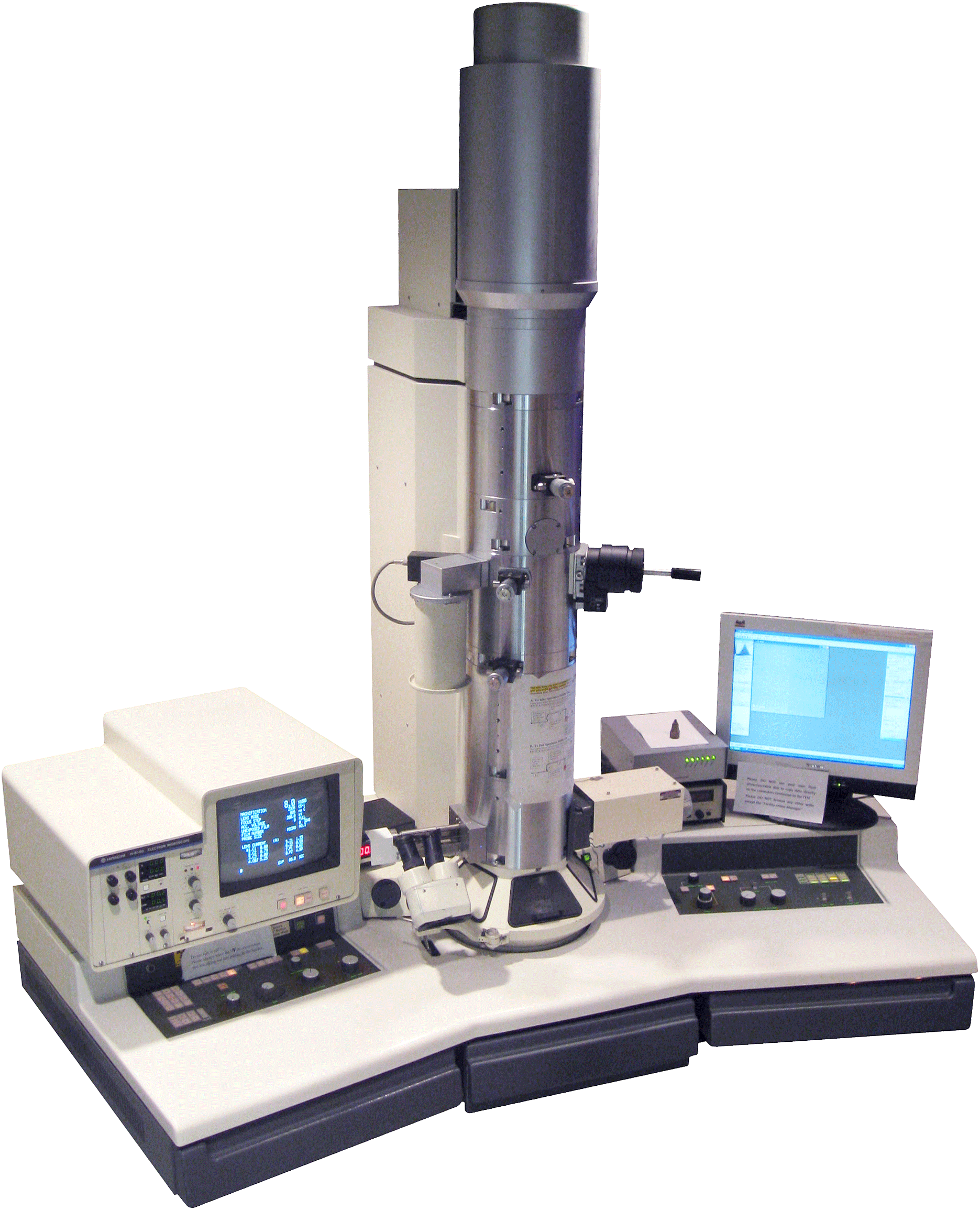
We use TEM Hitachi H‐7650 at 75kV to observe our origami.
Equipment
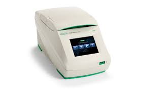
Thermal cycler
www.bio-rad.com
Thermal cycler is normally used for polymerase chain reactions (PCR) through the variation of temperature. We
use thermal cycler for folding reaction of NanoNeedle through thermal annealing.
Transmission electron microscopy (TEM)
www.nuance.northwestern.edu
Transmission electron microscopy allows a focused electron beam to transmit through an ultra-thin sample and
detect the electron scattered on the sample. We use the transmission electron microscopy to check the size and 3D structure of NanoNeedle.
Orbital shaking incubator and 37˚C, 5%CO2 incubator
www.labtech.in
For incubation of E. coli
orbital shaking incubator provides a constant shaking of samples; while 37˚C, 5% CO2 incubator provides a
suitable temperature as the in vivo environment.
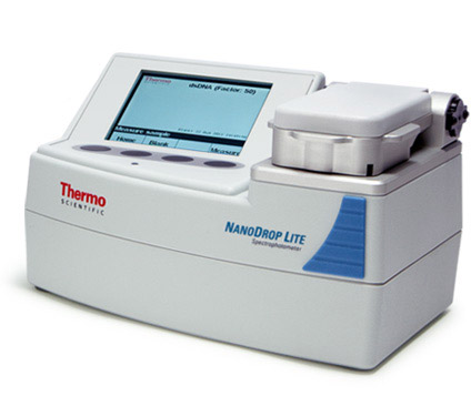
Nanodrop
www.nanodrop.com
Nanodrop is a supermicro-spectrophotometer to test the concentration of DNA. We use Nanodrop to determine the
concentration of the plasmid we extracted from the E. coli of midi-prep.
Flow cytometer
www.zmbh.uni-heidelberg.de
Flow cytometer uses the angle of scattering of fluorescent beam to separate particles of different properties.
We use flow cytometer to measure the GFP signal of E. coli incubated with NanoNeedle in functional test.
Centrifuge
www.directindustry.com
Centrifuge is a common equipment to purify or concentrate the sample. centrifuges work by the sedimentation
principle, where the centripetal acceleration is used to separate substances of greater and lesser density.<\p>
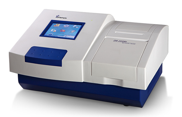
ELISA reader
ringbio.com
ELISA reader is a technique for the enzyme-linked immunosorbent assay (ELISA), a test using antibodies and color change to identify a substance. We use ELISA reader in MTT Assay to test the survival rate of HT29 cell line
after incubated with the NanoNeedle.
Cell counter
http://spectralab.net/mythic-18-hematology-cell-counter-human/
Cell counter counts cells alive and death cells respectively and determines the cell viability. We use cell counter to count the cells we have before we start to culture the human cells of colorectal cancer using in
cytotoxicity test.
Gel Electrophoresis
Gel electrophoresis allows the DNA segments in different lengths to be separated through agarose gel matrix, and clearly visualized under a UV transilluminator after staining with EtBr. We use gel electrophoresis to test the best concentration of MgCl2 for the folding environment, check whether the T7-GFP plasmid is digested completely by PstI and NanoNeedle is well folded.


